
Vascular Conditions
May-Thurner syndrome (MTS)
May-Thurner Syndrome (MTS) is a condition where a vein in your left leg gets squished by an artery in your right leg. This squeezing can cause swelling and discomfort in your leg. Sometimes, it can even lead to blood clots called deep vein thrombosis.
At PIIT our specialists doctors offer a simple treatment. They place a tiny tube called a stent in the squeezed vein to help it stay open. This can relieve the pressure and make you feel better.
How is the procedure performed at PIIT?
The procedure usually takes about an hour. You’ll be given light sedation, but you will be awake. A special doctor at PIIT will make a small cut near your neck or groin. They will put a thin tube called a catheter through the cut and into a vein. Then they will use a special machine called an ultrasound to check the vein. Sometimes doctors also use another machine called an intravascular ultrasound (IVUS) to check more. If there’s a problem, they put in a small metal cage, called a stent, to help fix it. This stent keeps the vein open. After this, the patients blood flow should get back to normal.
Deep Vein Thrombosis (DVT)
Deep Vein Thrombosis (DVT) is when blood clots form in deep veins, usually in the legs. These clots can travel to the lungs, causing a condition called Pulmonary Embolism (PE). This is very serious and can be life-threatening.
DVT is quite common and affects many people each year around the globe. There are different reasons why someone might develop DVT, like obesity, surgery, or being inactive for a long time. Sometimes, the cause is unknown. Symptoms of DVT include pain, swelling, and redness in the affected area. Sometimes there are no symptoms at all. Over time, DVT can lead to complications like chronic venous insufficiency, where the veins don’t work well, and post-thrombotic syndrome, which causes pain, swelling, and sores in the affected area. These complications can make it hard to walk or do daily activities.
How are DVT conditions treated at PIIT?
Doctors at PIIT can suggest one of the following treatments based on the evaluation of your condition.
- Clot busters are medicines that help break up blood clots. Your doctor might give them directly into the clot using a special therapy called Catheter Directed Thrombolysis (CDT). This treatment, done by an Interventional Radiology specialist, can improve symptoms and complications of DVT in carefully evaluated patients.
- Another method to remove clots is using small devices, smaller than a pen’s width, that can be placed directly into the vein by the same specialist. These devices help remove the clot, allowing normal blood flow again.
- Sometimes, a filter is placed in a large vein called the vena cava to stop clots from reaching the heart and lungs. This filter is called an inferior vena cava filter.
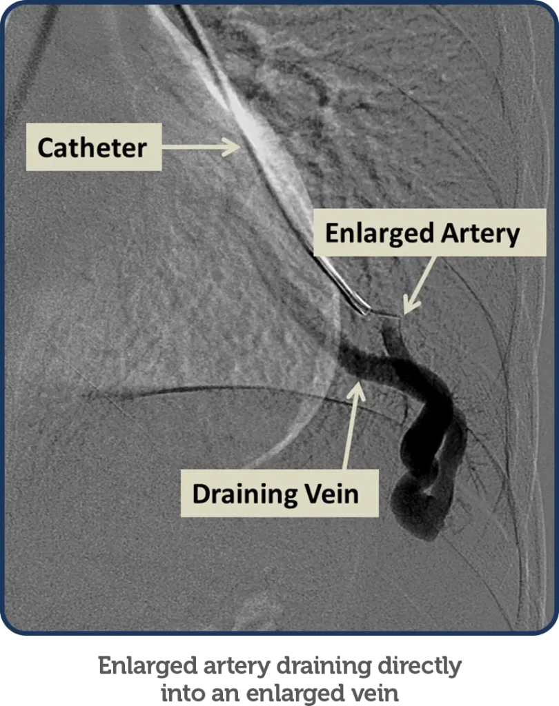
Arteriovenous Malformation (A.V.M) Embolization
What is (A.V.M) Arteriovenous Malformation ?
AVM is an abnormal tangle of blood vessels linking arteries and veins that interferes with regular blood flow and oxygen circulation.
Why this procedure is recommended?
This procedure is recommended in the following cases:
- Before surgery or radiation to reduce hemorrhage.High-output cardiac failure (heart failure).
- Chronic venous hypertension (increased pressure inside your veins).
- Lesions at a limb or life-threatening condition.
- Disabling pain.
- Functional disability.
- Cosmetic deformities.
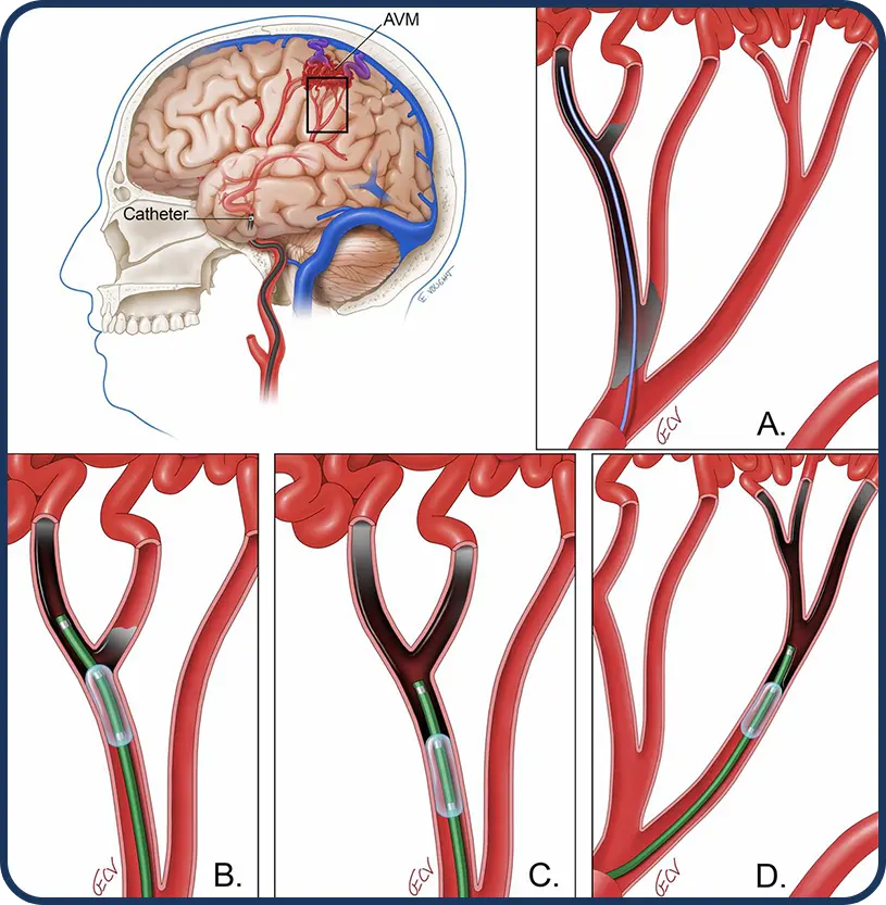
How is this procedure performed?
- The procedure begins with the doctor prepping the area over your groin with iodine soap.
- Local anesthesia will be injected into the skin at the insertion site. You may feel some stinging at the site for a few seconds after the local anesthetic is iniected.
- After the area is numb, a contrast dye is injected.
- Under image guidance, a needle will be inserted into the artery then a guidewire is placed through the needle into the artery
- A small catheter is guided through the needle into the artery supplying the AVM.
- A series of x-rays are taken to confirm that the catheter is placed in the correct position.
- Each AVM is then accessed using a catheter and blocked with small fibered coils or plugs.
Pulmonary Arteriovenous Malformation(AVM) Embolization
Malformations of the artery and vein are know as arteriovenous malformations. When they develop in the lung, they are termed pulmonary arteriovenous malformations (PAVM). PAVM Embolization is a procedure that blocks one or more pulmonary arteries that are supplying the malformation.
How is this procedure performed?
- The procedure begins with the doctor prepping the area over your groin with iodine soap.
- Local anesthesia will be injected into the skin at the insertion site. You may feel some stinging at the site for a few seconds after the local anesthetic is iniected.
- After the area is numb, a contrast dye is injected.
- Under image guidance, a needle will be inserted into the artery then a guidewire is placed through the needle into the artery
- A small catheter is guided through the needle into the pulmonary arteries.
- A series of x-rays are taken to confirm that the catheter is placed in the correct position.
- Each AVM is then accessed using a catheter and blocked with small fibered coils or plugs.
Arterial Leg Angioplasty
What is Leg Angioplasty?
What is a Stent?
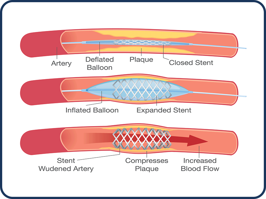
Angioplasty is a procedure to open narrowed or blocked blood vessels that supply blood to your legs.
A stent is a metal mesh tube that can be inserted into a narrowed artery. It acts as an internal support framework to keep the artery open by continuing to press plaque back against the artery wall.
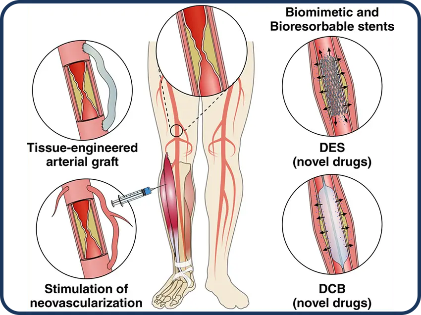
Why is this procedure recommended?
This procedure is recommended if you have peripheral artery disease (PAD), which can cause:
- Chronic pain in your legs.
- Heavy feeling in your legs.
- Limitations to vour daily activities.
- Wounds on your legs or feet that do not heal.
How is this procedure performed?
The procedure begins with the doctor prepping the area over your leg with iodine soap.
- Local anesthesia will be injected into the skin at the insertion site. You may feel some stinging at the site for a few seconds after the local anesthetic is injected.
- After the area is numb, a contrast dye is injected into the vein.
- A small tube is then placed into the blood vessels
- Under image guidance, through a small tube, a catheter is placed with a small balloon on the end of it.
- The balloon will be placed where the narrowing or blockage is and inflated a few times to open up the narrowed area
- A stent may then be inserted along with the balloon catheter. It expands when the balloon is inflated. The stent is left there to help keep the artery open. When the procedure is complete, the catheter is removed and the pressure is applied to stop any bleeding.
Bronchial Artery Embolization For treating bleeding in Lungs
What is Bronchial Artery Embolization?
Bronchial artery embolization is a minimally invasive procedure that stops bleeding by reducing blood supply to the damaged blood vessels in the respiratory system.
Why this procedure is recommended?
This procedure is recommended in the treatment of hemoptysis (Blood is coughed up from lungs or airway), which occurs due to:
- Chronic inflammation (inflammation of prolonged duration)
- Tuberculosis (Infection that mainly affects the lungs and is highly contagious, can also spread to other parts of your body)
- Aspergillosis (infection caused by a fungus that usually affects the lungs)
- Cystic fibrosis (an inherited disorder that causes the production of thicker mucus than usual which is difficult to cough out)
- Bronchiectasis (a disorder where the airways of lungs become widened that leads to the building up of excess mucus)
- Neoplasms (cancer of lungs)
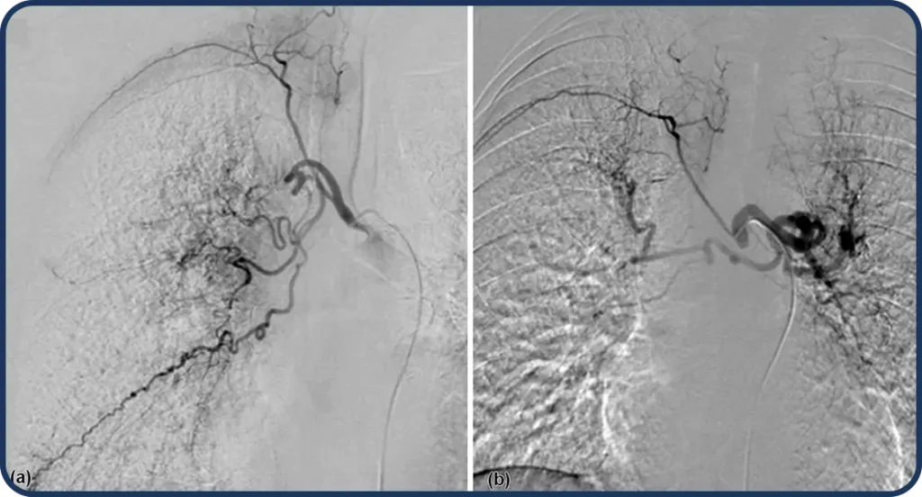
How is this procedure performed?
- The procedure begins with the doctor prepping the area over your groin with iodine soap.
- Local anesthesia will be injected into the skin at the insertion site. You may feel some stinging at the site for a few seconds after the local anesthetic is injected.
- After the area is numb, a contrast dye is injected.
- Under image guidance, a needle will be inserted into the artery then a guidewire is placed through the needle into the artery.
- The needle is taken out, allowing a fine plastic tube to be placed over the wire.
- A series of x-rays are taken to confirm that the catheter is placed in the correct position.
- The doctor will then inject small particles into the bleeding blood vessel, which stops the bleeding.
- After the procedure, the catheter will be removed.
Renal Artery Stenosis
Renal artery stenosis is when one or more arteries carrying blood to your kidneys become narrow. This usually happens because of atherosclerosis, which can hurt your kidney tissue. It can also lead to high blood pressure in your body, known as renovascular hypertension (RVH).
Risk factors for renal artery stenosis include age, smoking, high blood pressure, high cholesterol, diabetes, obesity, and not being active. Usually, there are no obvious signs, but sudden or worsening high blood pressure might be a clue. Doctors might find it while testing for other health issues.
The main cause is atherosclerosis, where plaque builds up and blocks the artery. Another cause is fibromuscular dysplasia, which affects artery muscle growth and can lead to early high blood pressure. Sometimes, it’s due to inflammation or growths pressing on the arteries.
Complications can include kidney failure, swollen legs, and difficulty breathing due to fluid buildup in the lungs.
How is Renal Artery Stenosis Treated at PIIT?
Renal Artery Stenosis is treated by a procedure called “Angioplasty”. It uses a balloon-tipped catheter to open a blocked blood vessel and improve blood flow. The doctor may place a metal mesh tube called a stent inside the blood vessel to help keep it open. This is the most common procedure if treatment is indicated. Learn more in the Angioplasty & Stenting procedure
Angioplasty & Stenting
What is Angioplasty?
What is Stenting?
Angioplasty is a procedure used to open narrowed or blocked blood vessels in your body.
A stent is a metal mesh tube that can be inserted into a narrowed artery. It acts as an internal support framework to keep the artery open by continuing to press plaque back against the artery wall.
Why is this procedure recommended?
This procedure is commonly used to treat conditions that narrow or block blood vessels and interrupt blood flow. These conditions include:
- Coronary artery disease (narrowing of the arteries that carry blood and oxygen to the heart muscle)
- Narrowing of the large arteries due to hardening of the arteries or atherosclerosis (narrowing of the large arteries due to build-up of cholesterol and other fatty deposits)
- Peripheral artery disease (PAD), a narrowing of the arteries in the legs or arms.
- Carotid artery stenosis, a narrowing of the neck arteries supplying blood to the brain.
- Narrowing or blockage in the veins in the chest, abdomen, pelvis, arms and legs.
- Renal vascular hypertension, high blood pressure caused by a narrowing of the kidney arteries.
- Narrowing in dialysis fistula or grafts (fistulas and grafts are artificial blood vessel connections doctors use in kidney dialysis.
Angioplasty is generally used when these connections become narrowed or blocked. Stenting may also be needed in some cases.
How is this procedure performed?
The procedure begins with the doctor prepping the area over your artery (in your wrist or
your groin) with iodine soap.
- Local anesthesia will be injected into the skin at the insertion site. You may feel some stinging at the site for a few seconds after the local anesthetic is injected.
- A sheath (plastic tube) will be inserted into the blood vessel.
- The catheter will be threaded into the arteries. Once the catheter is in place, contrast dye will be injected through the catheter into your arteries in order to see the narrowed areas.
- A series of rapid x-ray images of the arteries will be taken. When the doctor locates the narrowed artery, the catheter will be advanced to that location and the balloon will be inflated to open the artery. You may have some discomfort at this point because the blood flow is temporarily blocked by the inflated balloon.
- A stent may then be inserted along with the balloon catheter. It expands when the balloon is inflated. The stent is left there to help keep the artery open.
- When the procedure is complete, the catheter is removed and the pressure is applied to stop any bleeding.
Bleeding Embolization
What is Bleeding Embolization?
Embolization is a minimally invasive treatment that blocks one or more blood vessels or abnormal vascular channels. Embolization for bleeding is usually performed for patients who experience excessive or prolonged bleeding.
Why this procedure is recommended?
How is this procedure performed?
Bleeding embolization is usually performed to prevent abnormal bleeding in any part of the body. Some common health problems that can be treated using embolization are listed below:
- Bleeding that results from a traumatic injury, such as abdominal or pelvic injuries caused by a motor vehicle accident.
- Bleeding from the gastroduodenal artery (GDA) due to gastrointestinal tract lesions such as a peptic ulcer.
- Bleeding from vascular malformations. For example, abnormal vascular channels can form in between arteries and veins in the circulatory system of the lungs. (BAE)
- Bleeding from splenic artery due to hypersplenism or splenic malignancy
- Bleeding from the lower gastrointestinal tract (LGIB) due to colonic polyp or diverticular disease
- The procedure begins with the doctor prepping the area over your groin, wrist or other specific insertion site with iodine soap
- Local anesthesia will be injected into the skin at the insertion site. You may feel some stinging at the site for a few seconds after the local anesthetic is injected
- After the area is numb, a contrast dye is injected
- Under image guidance, a catheter is inserted through the skin to the treatment site.
- An embolic agent is then injected through the catheter and a series of x-rays are taken to ensure the procedure was a success and theblood flow in the target blood vessel or abnormality has been reduced or blocked as planned.
- After the procedure is completed, the catheter is removed.
Limb Salvaging
What is Limb Ischemia (C.L.I)?
The blood flow to a leg or arm is reduced over a long period. It happens because of narrow or blocked arteries that carry blood to the limb. This can cause pain, difficulty in walking, and wounds that are slow to heal. However, with appropriate Intervention, the symptoms of C.L.I. can be treated and the quality of life can be improved. Chronic limb ischemia is a condition that can affect a significant number of people. The important thing is to seek help from a doctor if there are any problems. When people get treatment, they can feel much better, and their leg or arm can work more easily
Common Symptoms
- Pain or discomfort in your leg or arm, especially during physical activity or when using the affected limb. The pain may subside with rest but return when you resume activity.
- Muscle weakness or fatigue of the affected limb
- Coldness and numbness at the affected limb
- Skin changes on the affected limb may appear pale, bluish, or have a shiny or thin appearance. It may feel dry, and you may notice hair loss or slow hair and nail growth in the affected area.
- Slow wound healing on the limb
- Non-healing ulcers often located on the feet or lower legs
- Gangrene may occur, leading to blackened, discolored, or foul-smelling skin.
- It’s important to note that symptoms of CLI can progress gradually over time, and not everyone will experience all of these symptom
Saving the Limbs
The doctors at PIIT use a tiny tube called a catheter to reach the blocked area in the leg or arm. They use small balloons or stents (mesh tubes) to open up the narrowed or blocked arteries. This helps to improve blood flow and reduce pain. It’s like fixing a clogged pipe so that water can flow freely again.